


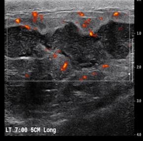
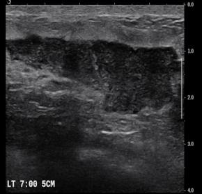
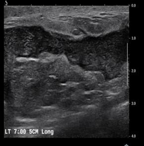
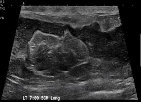
[Breast] 35 /F, Palpable mass, left with redness
|
|
|---|---|
| Subspecialty | Breast |
| Classification | Inflammation |
| Difficulty | For Student, For resident |
| Modality | US |
|
|
|
| Questions | What is your diagnosis? Go to answer |
| ANSWER | |
|---|---|
| [Breast] 35 /F, Palpable mass, left with redness | |
| Questions | What is your diagnosis? |
| Diagnosis | Idiopathic granulomatous mastitis |
| Answer | |
| Comments | Idiopathic granulomatous mastitis (IGM) is a rare benign inflammatory breast entity characterized by lobulocentric granulomas. IGM has a persistent or recurrent disease course and affects parous premenopausal women with a history of lactation. It has also been associated with hyperprolactinemia. The most common clinical sign is a palpable tender mass. However, the nonspecific manifestations and varied demographic features of this condition, as well as the other similar-appearing and superimposed breast entities, pose substantial diagnostic challenges. Entities with similar manifestations include inflammatory breast cancer (IBC), infective mastitis, foreign body injection granulomas, mammary duct ectasia, diabetic fibrous mastopathy, and systemic granulomatous processes. The strategy for imaging IGM depends on patient age, clinical manifestations, and risk factors. Targeted ultrasonography, mammography, and less commonly, magnetic resonance imaging have proven to be useful for imaging evaluation. Core-needle biopsy, with or without fine-needle aspiration for cytopathologic examination, and culture analysis are usually required to exclude IBC and other benign inflammatory breast processes. Patients with IGM have an excellent prognosis when they are appropriately treated with oral steroids or second-line immunosuppressive and prolactin-lowering medications. However, surgical excision may be an option for patients in whom medication therapy is unsuccessful. Imaging surveillance can be offered to patients with incidentally encountered IGM or mild symptoms. Clinical suspicion for this rare disease and the breast imager’s prompt diagnosis can lead to an improved patient outcome. The purpose of this article is to review the imaging manifestations of IGM in a multimodality case-based format and to describe relevant clinical and imaging-based differential diagnoses. The associated pitfalls, epidemiologic and histopathologic factors, clinical manifestations, natural course, and management of IGM also are discussed. |
| References | Radiographics . Mar-Apr 2018;38(2):330-356. doi: 10.1148/rg.2018170095. |
| Keywords | Mastitis, Granulomatous |
![]()
[04158]서울시 마포구 마포대로 53 마포트라팰리스 A동 304호
- 사업자등록번호 : 229-82-00887
- 대표자: 이재영
Copyright©by Korean Society of Ultrasound in Medicine
